One would think internet should be full with Electron Microscope (EM) images of COVID inside a human cell. Just several, inconclusive. I fail to see in this image the bronchitis coronavirus virions forming inside of a cisterna. Even the (sub)title of the image says: "assembling in a Golgi region"

Spent most of the morning searching for things like what type of human cells are attacked first by COVID and how COVID multiplies after taking over those cells. However i could not find EM images with those cells. All i could find was this. Here, an Endothelial Type II cell is shown sitting at the bottom (top in image) of an alveolar sack and its internal structure which includes a Golgi apparatus.

It is within this type of cell the exchange of gases between air and blood occur. Oxygen passes from air to blood and CO2 back in the air.
Current theories say COVID is packed (assembled) inside of organelles of human cells known as Golgi apparatus, more precisely in a structure of this organelle called cisterna. Prior entering the cisternae, in the intermediate endothelian reticulum or ER, ribosomes, enzyme like structures made of two proteins which normally produce proteins, one of the main functions of most cells, provides proteins for the viruses. Normally specific parts of RNA (which is the same for every cell of our body) from cell's nucleus is used by ribosomes as a blue print for cell specific proteins, then proteins made by ribosomes are evacuated when vesicles form at the end of Golgi apparatus cisternae and those vesicles are transported at the surface of the cell and evacuated in the intracellular space to do whatever.
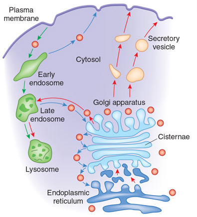
While i understand that viral RNA can be used instead of nucleus RNA in ribosomes to produce proteins (though i believe each type of cell has its own unique type of ribosome, which can have a limited "range" or what type of proteins can produce), i fail to understand how the Golgi apparatus and ER functions are changed (by the virus) to also produce membranes and envelopes.
I could not find an electron microscope image of the Golgi apparatus with cisternae for an Endotelial Type II cell, all i could find was one for white cells. The scale bar show that the thickness of cisternae and vesicles are smaller than 100 nm, the average size of a coronavirus, and that without the spike protein.


Ok it wouldn't be fair if i didn't put here what i found later. Vero is a type of "standard" lab grown cell coming from a single lineage descending from the liver cells of a specific monkey. Shown is a herpes virion building up at the end of a much smaller cisterna inside a vero cell. Looking at the image makes me think the cisterna receives the virion RNA from nucleus and provides the membrane, envelope, the M, S and spike protein, like it actually had this functionality and viruses could actually play an evolutionary role. But then in the explanation i see the word "budding" (c). Then i see several virions inside a single vacuole ready to be lifted at the surface of the cell.

From now on it becomes confusing, cause if you asked google, it says viruses are assembled inside golgi, if you follow this article, it's a different story. In both cases it seems obvious to me that the virus does not contain enough information to "hijack" or "reprogram" the cell into doing all this, but the cells are already adapted to do this since billion years ago to do it, like it had this functionality and viruses could play key roles in evolutionary process. Lipid membrane, envelope is provided by the ER and golgi, which pre-exists in host's cells.

One thing to mention here. When i was thinking that the cell itself could actually have this functionality and especially when i used the word "evolutionary", the man upstairs started to yell as much as one could yell obscenities in Spanish or Portuguese. Can not say i actually got scared and stopped reading and following ideas especially because apartment is filled with smoke from the walls. Got to finish or even re-write this post later. I would imagine the catholic point of view on this. God could not have created a human being (in its own resemblance) that produces viruses to infect others. Then i remembered the logo of the management firm of the apartment complex, and its resemblance to Dominican Province of the Philippines logo.
And one more thing. Here is a crop from last EM image.

No comments:
Post a Comment
Friendly comments welcome
Note: Only a member of this blog may post a comment.